Suppression Tests
Suppression is common in patients with amblyopia and strabismus (lazy eye). Suppression tests give the clinician an accurate measurement of what the patient is able to see centrally. These tests can be utilized throughout a therapy treatment plan to reassess how the patient is progressing in reducing suppression. With time and vision therapy, the patient can reduce their suppression significantly and even eliminate it. Suppression occurs mostly in the patients central vision, the tests of suppression will test the patients suppression in the central vision.
What is suppression?
Suppression essentially refers to the brain ignoring the visual signal provided by one eye. Let's start with a normal pair of eyes. When the eyes are aligned and pointing towards the same object in space and have an equally clear image, the brain uses small cues to determine the depth, size, shape, etc. of the object and combines the images of each eye together to see.
If one eye has a very blurry image (amblyopia, specifically refractive amblyopia due to an unequal glasses prescription) the brain struggles to combine the images of each eye together into a single image. To compensate, the brain suppresses or ignores visual input from the blurry eye. This same concept occurs if one eye is turned (strabismus or squint). In this example, the brain sees multiple images, which would be confusing. Again, the brain suppresses (ignores) visual input from the deviating eye and utilizes input from the fixating eye.
At its core, suppression is the visual system's method of compensating or adapting for a defect. To avoid the confusion of a blurry image superimposed on a clear image, or seeing two of one image in space, the brain simply ignores the abnormal image. Suppression is rather easily "learned" by the brain at an early age when the visual system has a higher degree of neuroplasticity. In contrast, a patient that suddenly acquires a strabismus or squint later in life may have trouble suppressing and constantly see double.
For amblyopic patients, a critical issue stems from long-term suppression. Suppression can be difficult to "unlearn". One of the key components of visual rehabilitation for patients that have amblyopia or strabismus is to teach the brain to use both eyes together. Tests of suppression check for the presence of suppression and, in some cases, quantify the amount of suppression the visual system is experiencing.
Tests of Suppression
Worth Dot / Four Dot Test
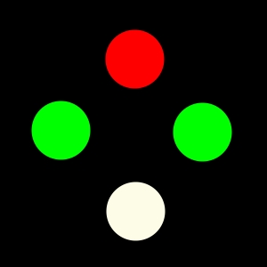
The four dot test uses red-green glasses and a flashlight or other light source with red, green, and a single white dot filter on it. The patient fixates on the light and reports the total number of lights or shapes seen. It is important that the patient has both eyes open during the test and that they keep them open (with blinking of course) while performing the test. This ensures an accurate result of the Worth Dot/ Four Dot test. The test itself can take just a few moments, this is why it has become a favorite amongst the Neuro-Developmental Optometry community.
Image of the Worth 4 Dot Test

When the patient is reporting the total number of shapes, this can give the clinician an idea of which eye is dominating as well as how the eyes are positioned. Colors can be adapted for the color deficient community. When the colors are adapted, the patient would wear the colored glasses corresponding with the colors on the Worth Dot/ Four Dot test.
Striated Lens Test (Bagolini Lens Test)
Striated lenses are special lenses with no power or correct in them. Instead, these lenses have small lines (striations) that run at 45 degrees and parallel in one eye and 135 degrees and parallel in the other eye. The clinician uses a penlight and asks the patient to look directly at the light. The result for the patient is the projection of an X pattern of light (normal vision) or missing lines or pieces of a line missing (example / seen if suppression one eye or \ seen if suppression the other).
Interestingly enough, this test can also give an indication of the patients eye posture, subjectively. Some patients may see both diagonal lines, however, the lines may not intersect to make an X they may look more like an upside down V or not intersect at all.
4 Base-out Prism Test
Prisms have numerous applications in vision care. Prism functions by bending light, which alters where an image appears to be in space. The human visual system can often detect very small movements of images, so when a small amount of prism is applied in front of one eye, images appear to move slightly and the eyes make a quick adjustment. However, if one eye is suppressing, the small image movement goes unnoticed, and the eyes will not make an adjustment. This is very quick and helpful suppression check for even small eye turns (called microtropia).
When the clinician performs this test, the clinician is able to achieve a quick understanding of the patients eye movements in a short amount of time.
Brock String
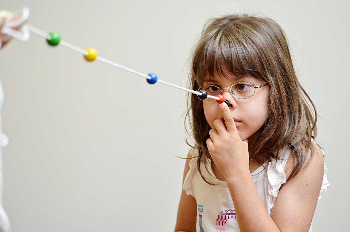
The Brock string is more commonly used as a training tool, but also has some value as a quick suppression check. The patient holds a string with different colored beads attached to it up to his nose. When focused on different beads, the patient should notice the string forms an X or V (depending on where the beads are on the string). This is using the concept of physiological diplopia - the string should appear doubled, but if a patient is suppressing, only one part of the string will be seen.

The Brock String also offers a bit of subjective knowledge to the patient of where their particular eyes are pointed in space. This can be very important for the recovery of binocular function for the patient. Letting the patient know where their eyes are pointed based on the Brock String incentivizes the patient to put their eyes in the "correct" position. The Brock String is a staple of vision therapy and can be used in a variety of ways outside of a suppression test.
The Vision Wiki
- Binocular Vision
- Vision Tests
- Suppression Tests
- Worth 4 Dot
- Tests of Stereopsis
- Cover Test
- Definitions
- Signs and Symptoms
- Blurry Vision
- Reading Problems
- Eye Strain
- Driving Problems
- Headaches
- Suppression
- Double Vision
- Motion Sickness and Car Sickness
- Pias Vision
- Visual Skills
- Visual Tracking
- Visual Fixation
- Stereopsis
- Depth Perception
- Visual Accommodation
- Visual Requirements for Baseball
- Visual Requirements for Pilots
- Reading
- Foundational Reading Skills
- Vision and Learning
- Fusion
- Convergence and Divergence
- Reading Skills
- Visual Processing
- Eye Problems
- Physiology of Vision
- Lazy Eye
- Lazy Eye Treatments
- Reading
- Fields of Study
- Research
- Glaucoma
- Virtual Reality
- Organizations
